
Time Stamps:
- 0:21 Introduction
- 1:39 Yapping Verteporfin: The importance in hair transplantation and hair follicle regeneration
- 7:01 Verteporfin Hair Transplant Results
- 14:13 Wound Healing Versus Regeneration
Clinical studies by Dr. Barghouthi and Dr. Bloxham suggest that using Verteporfin with FUE and FUT hair transplantation methods can lead to promising hair follicle regeneration and minimal scarring. Verteporfin, known for inhibiting Yes-associated protein and reducing scarring, shows potential in enhancing regenerative healing, but there are concerns about its dose dependency and the need to determine the optimal concentration for effective treatment.
Additionally, the DHT sensitivity of regenerated follicles remains a concern due to the influence of embryonic development on male pattern baldness. The precise depth of microneedling, crucial for maximizing Verteporfin’s effects, also needs further investigation to ensure the treatment targets the appropriate skin layers without causing undue harm. As research progresses, finding the right balance of dose, application method, and adjunctive treatments will be key to improving outcomes. While these findings are promising, they are part of emerging research and should be interpreted as supplementary to professional medical advice.
While this presents a potential breakthrough, it’s crucial to note that this is emerging research and not a substitute for professional medical advice.
- Clinical trials by Dr. Barghouthi and Dr. Bloxham indicate promising results in hair follicle regeneration and minimal scarring with Verteporfin and Hair transplantation methods FUE and FUT.
- Dr. Barghouthi: Clear signs of regrowth, minimal scaring
1. https://www.hairrestorationnetwork.com/profile/32181-drtbarghouthi/
2. https://www.hairrestorationnetwork.com/topic/64737-verteporfin-hair-regeneration-human-trial-dr-barghouthi-official-thread/page/47/#comment-744349 - Dr. Bloxham: Slow healing, signs of regrowth, minimal scaring

- Q: Could microneedling at a depth between 3mm to 3.5 mm in bald or thinning areas reactivate dormant hair follicles/regenerate new ones?
- 3mm to 3.5 mm is the extraction range for hair follicles in the scalp. The justification of microneedling at this depth is to induce a similar ‘depth of trauma’ to that which is experienced in hair transplantation.
1. Örs, S. (2017). Follicular unit extraction (FUE) hair transplantation. In In Tech eBooks. Follicular Unit Extraction (FUE) Hair Transplantation | IntechOpen - Here, after microneedling at this painful range verteporfin could either be injected into the scalp or somehow applied topically
Hypothetical Protocol with Verteporfin and Microneedling:
- 5AR Treatment: Scalp DHT must be reduced to produce conducive environment for regenerating hair follicles that MAY (high likelihood) be DHT sensitive.
- Why is this a high likelihood? The following is an article in The Guardian: ‘We’re not making new hairs, we’re rescuing’: could scientists reverse male pattern baldness? | Hair loss | The Guardian It talks about how male pattern baldness patterns could potentially be predicated from as early as embryonic development. Hair Follicles that are DHT sensitive could have a different cell linage.
- Preparation and Cleaning: Ensure the scalp is clean to reduce infection risks.
- Microneedling Procedure: Perform microneedling at a depth of 3mm-3.5mm to target the deeper layers of the scalp effectively.
- Application of Verteporfin: Apply Verteporfin topically or inject post-microneedling to inhibit YAP activity, enhancing follicular regeneration and minimizing scarring.
- Post-treatment Care: Follow a stringent aftercare regimen to avoid infections and monitor healing, acknowledging the slower regenerative process.
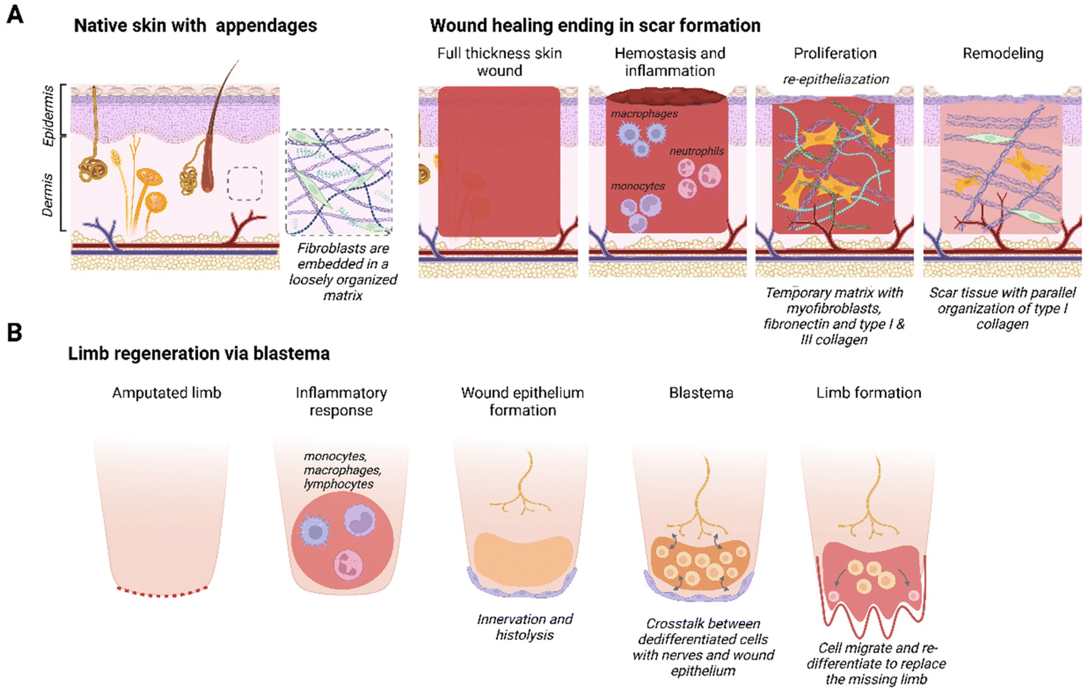
Overview of Healing in Mammals vs. Regenerative Species and the Role of YAP Inhibition
Healing in Mammals
Mammals, including humans, primarily heal through a process that results in scarring. When an injury occurs, the body’s immediate response is to close the wound, prevent infection, and restore integrity to the area. This involves a complex cascade of inflammatory responses, tissue formation, and remodeling, often resulting in scar tissue, which is fibrous and lacks the original tissue’s full functionality and appearance. The scarring process is quick and effective for survival but does not restore the original tissue’s architecture.

Erickson, J. R., & Echeverri, K. (2018). Learning from regeneration research organisms: The circuitous road to scar free wound healing. Developmental Biology, 433(2), 144–154. Redirecting
Regeneration in Other Species
In contrast, some species, such as certain amphibians and reptiles, can regenerate lost limbs or organs. This regeneration involves the re-growth of complex structures, including bone, muscle, and skin, to fully restore the lost part’s function and appearance. For example, axolotls can regenerate entire limbs, tail, heart, and other organs with perfect functionality and without scarring. The process involves dedifferentiation of cells, proliferation, and re-differentiation, meticulously reconstructing the lost part.
Avila-Martinez, N., Gansevoort, M., Verbakel, J. M. A., Jayaprakash, H., Araújo, I. M., Vitorino, M., Tiscórnia, G., Van Kuppevelt, T. H., & Daamen, W. F. (2023). Matrisomal components involved in regenerative wound healing in axolotl and Acomys: implications for biomaterial development. Biomaterials Science, 11(18), 6060–6081. Matrisomal components involved in regenerative wound healing in axolotl and Acomys: implications for biomaterial development - Biomaterials Science (RSC Publishing)
In the paper “Inhibiting Yes-associated protein prevents scarring and promotes regeneration in a large animal model of wound repair,” Heather E. Talbott and colleagues demonstrated that a single local administration of Verteporfin following injury significantly reduced scarring and induced regenerative healing in red Duroc pigs. Notably, these pigs are a close analog to human skin in terms of thickness and are often used in dermatological research, making the findings particularly relevant. The successful regeneration of scarless skin and hair follicles in these pigs points to the potential of Verteporfin to prevent scarring and regenerate hair follicles in humans as well.

Talbott, H. E., Griffin, M. F., Mascharak, S., Guardino, N. J., Abbas, D., Lavin, C., Guo, J. L., Spielman, A. F., Cotterell, A., Parker, J. B. L., Januszyk, M., Lorenz, H. P., Wan, D. C., & Longaker, M. T. (2022). Inhibiting Yes-associated protein prevents scarring and promotes regeneration in a large animal model of wound repair. In The 23rd Annual Emile F. Holman Lecture in Surgery and 13th Annual Resident Research Day (p. 28). Stanford University School of Medicine Department of Surgery. Retrieved from https://surgery.stanford.edu/content/dam/sm/surgery/documents/HolmanLectureBooklet_2022%20FINAL.pdf
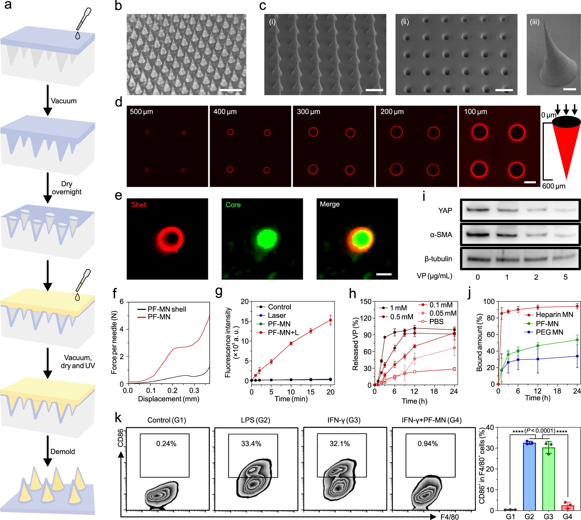
The paper titled “Scarless wound healing programmed by core-shell microneedles,” authored by Ying Zhang et al. and published in Nature Communications in 2023, provides further insights. It demonstrates the remarkable healing effects of Verteporfin on mice, where treated skin areas not only heal effectively but also closely resemble unaffected skin in appearance and functionality, indicating the broad potential of Verteporfin in promoting regenerative healing across species.
Zhang, Y., Wang, S., Yang, Y. et al. Scarless wound healing programmed by core-shell microneedles. Nat Commun 14, 3431 (2023). Scarless wound healing programmed by core-shell microneedles | Nature Communications
a. This is a diagram showing how the microneedle patch is made. The process starts with a mold that has sharp points (like an ice cube tray with spikes). A material is poured into the mold, vacuum is applied to remove air bubbles, and it’s left to dry. Then, it’s exposed to UV light to solidify, and finally, the finished product is removed from the mold.
b. A photo of the actual microneedle patch, which has many tiny needles on it. This is probably a small patch that could fit on your skin.
c. These are scanning electron microscope (SEM) images showing the needles of the patch from different views. The first one shows the side of the needles, the second shows the bottom, and the third is a close-up of a single needle.
d. These images were taken with a confocal microscope, which can look at slices of the sample at different depths. They are showing how deep the drug (labeled with a red dye) goes into the needle.
e. Here we see the needles have two parts: a core and a shell. The shell is marked with a red dye and the core with a green dye. When the two images are merged, it shows how the core is surrounded by the shell.
f. This graph shows the force needed to break the shell of the needle and the entire needle with the drug. It helps to understand how strong the needles are.
g. This graph shows the production of reactive oxygen species (ROS), which are important in the body’s defense and also for signaling when the needles are exposed to light.
h. This graph shows how the drug (Verteporfin or VP) is released from the needles over time in different conditions.
i. This is a Western blot, a technique used to detect specific proteins in a sample. It shows how the levels of certain proteins change in cells treated with different concentrations of the drug.
j. This graph shows how well different types of microneedles can bind to a protein called IFN-γ over time.
k. The last panel shows flow cytometry data, which is a way to measure characteristics of cells. This experiment was looking at how the needles affect the behavior of a type of immune cell called macrophages.
The text mentions that the needles are made of something called PF-MN, which probably refers to the materials used. The needles are designed to release their drug payload in response to something (like a change in the body’s chemistry), and the studies are showing how well they release the drug, how strong they are, and how they affect cells in the lab. The statistical significance indicates that the results are highly reliable (P < 0.0001).
Human Fetal Tissue Regeneration
Early in fetal development, humans have the ability to heal wounds without scarring. This is observed in the unique regenerative capacity of fetal skin, which can completely regenerate without scar formation until a certain point in gestation. The environment within the womb, including high hydration and specific growth factors, along with a distinct cellular response, allows for this scarless healing. As development progresses, this ability diminishes, and postnatal wounds typically heal by scarring.

Hu, M.S., Maan, Z.N., Wu, JC. et al. Tissue Engineering and Regenerative Repair in Wound Healing. Ann Biomed Eng 42, 1494–1507 (2014). Tissue Engineering and Regenerative Repair in Wound Healing | Annals of Biomedical Engineering
Significance of YAP Inhibition
Yes-associated protein (YAP) is a key regulator within the Hippo signaling pathway, crucial for controlling organ size, cell proliferation, and apoptosis. It’s also significantly involved in wound healing and tissue regeneration. In the context of healing, YAP influences the behavior of fibroblasts—cells that play a critical role in wound repair and the formation of scar tissue. When YAP is active, fibroblasts are more likely to create the fibrous, collagen-rich tissue that leads to scars. Inhibiting YAP can reduce this scarring tendency and encourage a more regenerative form of healing.

Zhang, Y., Wang, S., Yang, Y. et al. Scarless wound healing programmed by core-shell microneedles. Nat Commun 14, 3431 (2023). Scarless wound healing programmed by core-shell microneedles | Nature Communications
Speculating on the Appropriate Amount of Trauma for Scarless Healing
Understanding Trauma and Scarring: In wound healing, the amount of trauma sustained by tissue can significantly influence the healing outcome. A delicate balance exists between sufficient trauma to trigger the body’s natural healing mechanisms and excessive trauma leading to scarring. The body’s response to injury involves a complex interplay of cellular signals, structural changes, and inflammatory responses, all of which contribute to either regenerative healing or scar formation.
Role of En1 and YAP in Healing
Research by Shamik Mascharak and colleagues in “Preventing Engrailed-1 activation in fibroblasts yields wound regeneration without scarring” highlights the significance of Engrailed-1 (En1) as a mechanoresponsive regulator in fibroblast activation. When En1 is activated, typically in response to trauma, it guides fibroblasts to repair the wound in a way that often leads to scarring. YAP plays a pivotal role in this process by signaling the activation of En1, thus promoting the fibroblast response that leads to scar tissue formation. YAP inhibition prevents signaling to the EN1 gene, which is instrumental in directing fibroblasts during wound healing. By modulating this pathway, it’s possible to shift the behavior of fibroblasts from rapidly creating scar tissue to slowly rebuilding tissue structures in a way that more closely resembles the original, without scarring.
Determining the Optimal Trauma Level
In the context of microneedling combined with Verteporfin treatment, the goal is to induce a sufficient level of trauma to trigger the body’s repair mechanisms without crossing the threshold into excessive injury that results in scarring. Determining the optimal level of trauma requires a precise understanding of the skin’s tolerance and the intensity of the microneedling procedure. This involves considering the depth and density of needle penetration and the individual’s skin characteristics.
Introducing Verteporfin to Inhibit YAP
Once the appropriate level of trauma is administered via microneedling, Verteporfin is introduced as a YAP inhibitor. By inhibiting YAP, Verteporfin aims to prevent the activation of En1 and the subsequent fibroblast-driven scarring response. The hypothesis is that by modulating this pathway, the healing can shift towards regeneration, closely mimicking scarless fetal healing. The hope is that this approach will not only reduce scarring but also promote the regeneration of hair follicles in bald areas, leveraging the body’s inherent regenerative potential.

Shamik Mascharak et al. ,Preventing Engrailed-1 activation in fibroblasts yields wound regeneration without scarring. Science372,eaba2374(2021).DOI:10.1126/science.aba2374
Animal Studies and Verteporfin
Animal studies have been instrumental in understanding the potential of YAP inhibitors like Verteporfin in promoting regenerative healing. For example, research involving mice and other mammals has shown that applying Verteporfin to wounds can lead to less scarring and more regenerative-like healing. These studies are vital as they show the potential for YAP inhibitors to change the way wounds heal, potentially enabling a more regenerative process similar to what is observed in fetal tissue and regenerative species.
You can see the further elaboration in this science article. https://www.science.org/doi/full/10.1126/science.aba2374?rfr_dat=cr_pub++0pubmed&url_ver=Z39.88-2003&intcmp=trendmd-sci&rfr_id=ori%3Arid%3Acrossref.org

Shamik Mascharak et al. ,Preventing Engrailed-1 activation in fibroblasts yields wound regeneration without scarring.Science372,eaba2374(2021).DOI:10.1126/science.aba2374
Clinical Evidence from Dr. Barghouthi and Dr. Bloxham
Dr. Barghouthi and Dr. Bloxham have made significant contributions to the field through their clinical case reports and studies involving patients undergoing hair transplantation. It is reasonable to conclude that Verteporfin has led to improved outcomes, including reduced scarring and enhanced hair follicle regeneration, based on the evidence.
Questions on Verteporfin dosage and use
However, it remains unclear whether the effects of Verteporfin are dose-dependent. Moreover, it is yet to be determined if the hair follicles observed in Dr. Barghouthi’s and Dr. Bloxham’s case reports will demonstrate growth similar to that of native terminal hairs. I also have questions regarding the number of Verteporfin hair transplant rounds one can undergo before potentially experiencing a decline in hair follicle regeneration in the donor area, if such a decline occurs at all.
Scalp DHT Concern
Finally, scalp DHT will continue to be a concern. Even if we manage to regenerate follicles in bald areas, the potential for regrowth might depend on the concentration of scalp DHT. It would be prudent to reduce scalp DHT as much as possible to create a more hospitable environment, especially if the regenerated follicles share a similar cell-niche lineage with the former DHT-sensitive follicles that once inhabited the bald areas. This approach may increase the likelihood of successful hair regrowth in those areas.
Why is this a high likelihood? The following excerpt is from this article in The Guardian: ‘We’re not making new hairs, we’re rescuing’: could scientists reverse male pattern baldness? | Hair loss | The Guardian It talks about how male pattern baldness patterns could potentially be predicated from as early as embryonic development. Hair Follicles that are DHT sensitive could have a different cell linage.
In a paper published in May, Higgins described evidence that the hairline of a middle-aged man could be traced back to the earliest stages of embryonic development. Around the third week of an embryo’s life, cells form three layers called the ectoderm, mesoderm and endoderm. Most organs in the body contain cells that derive from just one of these lineages: the endoderm gives rise to the internal organs, the mesoderm becomes the muscle and connective tissues, and the ectoderm becomes the central nervous system. “Normally a tissue is one lineage, but the [skin] is a bit of an enigma,” says Higgins.
“The dermis [the skin’s lower layer] on the face is ectoderm and the dermis on your body is mesoderm, but the top of your head is not really known.” Higgins argues that male pattern baldness traces out the boundary between skin cells that have taken two very different paths during development. This, she says, could explain why the cells on only certain parts of the head are oversensitive to dihydrotestosterone. Kemp and colleagues are working to develop a test, based on the gene expression of dermal papilla cells, to establish whether the part of the scalp they come from is balding, destined to bald or will always retain its hair. “Ideally you want to be able to map the head,” he says. “We’re finding genetic differences between the hairs and we’re in the preliminary phases of doing that. (Devlin, 2023)
Devlin, H. (2023, October 1). ‘We’re not making new hairs, we’re rescuing’: could scientists reverse male pattern baldness? The Guardian . ‘We’re not making new hairs, we’re rescuing’: could scientists reverse male pattern baldness? | Hair loss | The Guardian
Based on the description provided in the paper by Higgins and colleagues, the high likelihood that regenerated hair follicles in bald areas would also be DHT sensitive stems from the developmental origins and lineage specificity of skin cells. Higgins suggests that male pattern baldness delineates the boundary between skin cells that have diverged along two distinct developmental paths. This implies that the predisposition for hair follicles to be sensitive to dihydrotestosterone (DHT) is fundamentally linked to their embryonic cell lineage—whether they originate from ectoderm or mesoderm cells.
If the regenerated hair follicles in bald areas are derived from or share a similar lineage to the original follicles that were sensitive to DHT, they would likely retain the same sensitivity characteristics. Since male pattern baldness is associated with the oversensitivity of certain scalp cells to DHT, particularly those in characteristically balding patterns, any new follicles that are regenerated or reconstituted from these same cell lineages would presumably exhibit similar DHT sensitivities. Essentially, the genetic and developmental blueprint that predisposes certain hair follicles to hair loss due to DHT sensitivity would still be present in the cells of those regions, influencing the characteristics of any newly regenerated follicles.